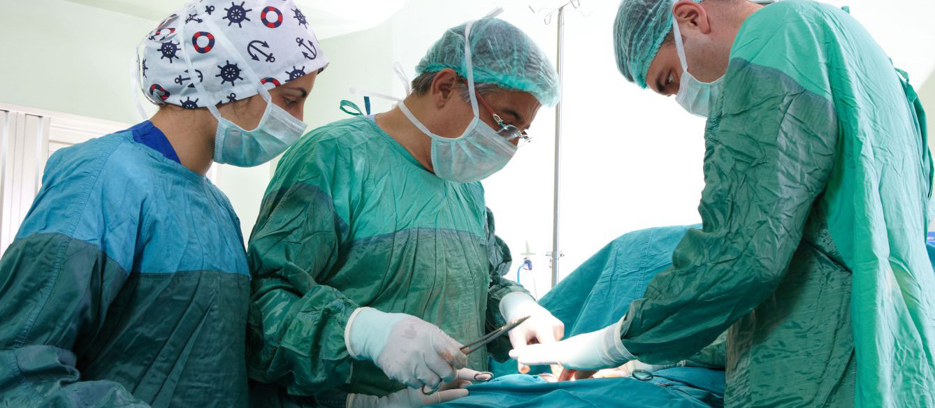
The device inserted into the cervix and uterine cavity to examine them is called hysteroscope, the procedure performed is called hysteroscopy.
In which areas is hysteroscopy used?
- Polyps and myomas: A myoma grown in the uterus is removed with hysterecopy. The same procedure is used for the treatment of a polyp grown in the uterus. Thus, the correct treatment is carried out by both removing the lesion and determining whether it is malignant or benign with patholocial examination.
- Asherman’s syndrome (intrauterine adhesions): Adhesions occur in the uterus as a result of surgeries and interventions performed. In medical terms, this condition is called Asherman’s syndrome. Hysteroscopy is used for the treatment of this disease. It opens the adhesions and restores them. Thus, a woman has regular menstruation and can conceive.
- Excessive bleeding: It is used to investigate the cause and make a correct diagnosis, if a woman’s menstrual bleeding increases a lot. Biopsy sample can also be taken thanks to the procedure performed.
- Recurrent miscarriage: Hysterectomy is performed to investigate why miscarriage occurs in women who continuously conceive and have miscarriage. If a woman, who had miscarriage, has miscarriage within the first 3 months and it constantly recurs, the cause is investigated. Solutions are found for such issues within the shortest time.
- Removal of intrauterine device: Intrauterine devices that cannot be removed under normal condition during gynecological examinations are removed very quickly and easily with hysterecopy.
- In the evaluation of IVF failure: It is used to investigate the causes when IVF fails.
- In the investigation of infertility causes: It is preferred to investigate the cause of infertility.
- Removal of intrauterine components: It is usually preferred in such cases.
How is hysteroscopy performed?
- It is not a procedure that requires hospitalization.
- It is performed in the operating room.
- It does not require anesthesia.
- The hysteroscopy method can be usually performed in the gynecological examination position and under spinal or general anesthesia.
- Sedative drug is given.
- The patient does not consume anything 6 hours before general anesthesia.
- The operation is first is cleaned with antiseptic.
- Clean and sterile drape is placed.
- The cervix is dilated up to 9 cm using a dilator.
- The hysteroscope is passed through this opening and placed into the uterus.
- A special fluid is given to clearly visualize the inside of the uterus. This liquid is re-cleaned. The blood and mucous secretions are cleaned and the vision becomes clear.
- Inside of the uterus is visualized with its camera. If there is a pathological problem after the examination inside of the uterus, operation is carried out.
- The material is sent to pathology after the operation.
- It produces result within 5 minutes or 1 hour depending on the type of surgery.
- The patient can be discharged a few hours or a day after the procedure.
- In addition, a small amount of bleeding and classic pain in the form of cramp occur for a day or two.
What are the risks of hysteroscopy method?
- Excessive bleeding,
- Injuries to the cervix,
- Intrauterine infection,
- Complications due to anesthesia.
